Figure 1.
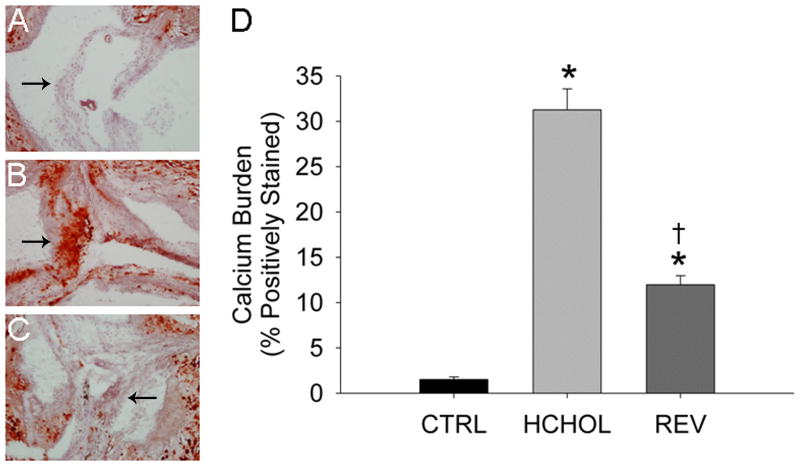
Effects of reducing plasma lipids on valvular calcium deposition (Alizarin Red staining) in A) control mice (CTRL), B) hypercholesterolemic mice (HCHOL), and C) “reversed” mice (REV). Images show valve cusps extending into the lumen of the aorta—arrows highlight corresponding/similar anatomical regions of the valve cusp in each image. Note that there is massive calcification of the aortic valve in HCHOL mice, which can be markedly reduced in a relatively short period of time by reducing blood lipids. D) Quantitation of histological data. * = p < 0.05 versus control mice, † = p < 0.05 versus hypercholesterolemic mice.

