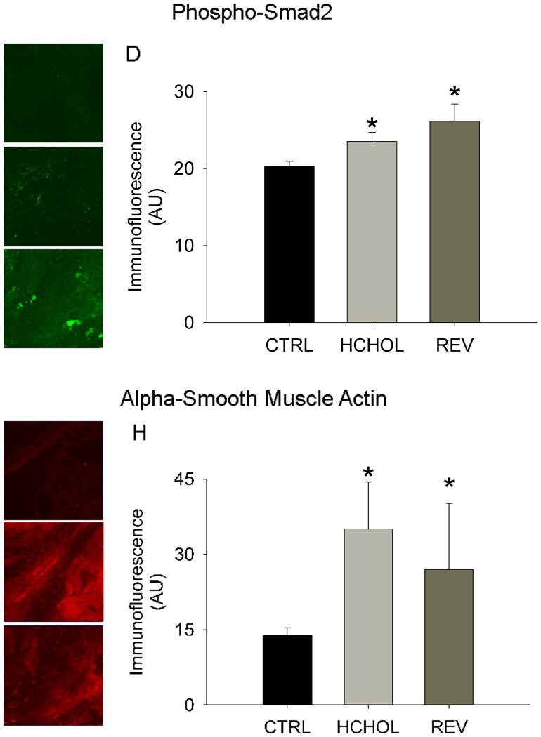
Figure 4.
Effects of reducing plasma lipids on pro-fibrotic/TGF-β signaling and myofibroblast activation. Phospho-Smad2 immunofluorescence (indicative of TGF-β signaling) in A) control mice, B) hypercholesterolemic mice, and C) “reversed” mice. D) Quantitation of phospho-Smad2 immunofluorescent data. Alpha smooth muscle actin levels (indicative of myofibroblast activation) in E) control mice (CTRL), F) hypercholesterolemic mice (HCHOL), and G) “reversed” mice (REV). H) Quantitation of α-SMA immunofluorescent data. * = p < 0.05 versus control mice, † = p < 0.05 versus hypercholesterolemic mice. Images were acquired using a 60x objective and include only valvular tissue near the base/attachment of the valve.

