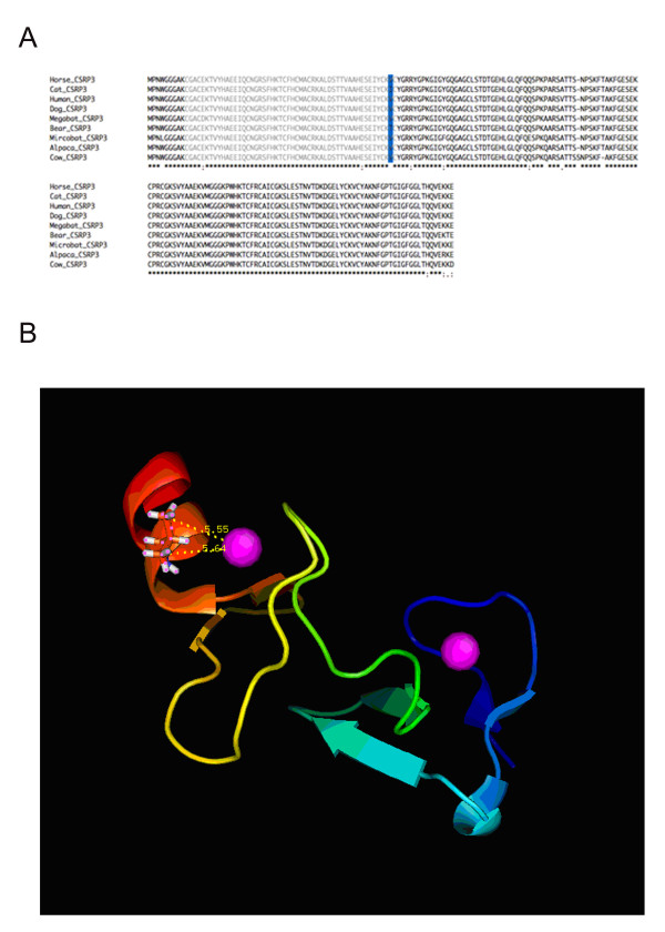
Figure 5.
Protein sequence alignment and 3D protein structure of CSRP3. (A) Multiple sequence alignment of full-length CSRP3 protein sequences in nine species. The LIM1 zinc-binding domain is marked with grey and the 60th site is highlighted in blue. The corresponding nucleotide sequence alignment is shown in Figure S5 (Additional file 6, Figure S5). (B) 3D protein structure of human CSRP3 LIM1 domain obtained from Protein Database (PDB id: 2o13). Two dash lines show distances between the zinc ion and the methyl groups of the valine residue.

