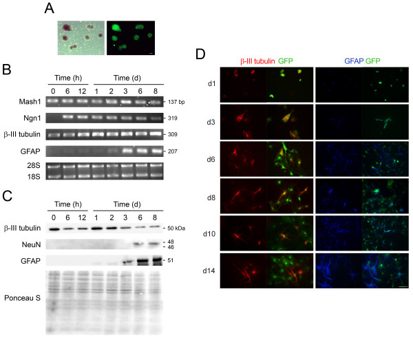
Figure 1.
Mouse NS cells have both neurogenic and gliogenic potential in vitro. Mouse NS cells were expanded in the presence of EGF and bFGF, and induced to differentiate as described in Materials and Methods. Cells were harvested for total RNA and protein extraction, or fixed at different time points and processed for immunostaining analysis. Neuronal differentiation was readily detected after incubation in differentiation medium, while glial production was only observed from day 3. A) Mouse NS cells constitutively expressing GFP were grown in suspension as neurospheres that increased in number and size with cell proliferation (phase contrast and GFP fluorescence images). B) Semi-quantitative RT-PCR analysis for selected markers of lineage commitment at successive time-points of mouse NS cell differentiation. Neurospheres (undifferentiated NS cells) collected before platting in differentiation medium were considered 0 time differentiation. Data was normalized to 28 S. C) Representative immunoblots showing protein expression of neuronal (β-III tubulin and NeuN) and glial (GFAP) markers during mouse NS cell differentiation. Ponceau S was used as loading control. D) Mouse NS cells labeled with anti-β-III tubulin and anti-GFAP antibodies, to visualize neurons and glial cells, respectively. Representative immunostaining images were from at least 3 independent experiments. Scale bar: 50 μm. Ngn1, neurogenin1; GFAP, glial fibrillary acidic protein; NeuN, neuronal nuclei; h, hours; d, days.

