Figure 4.
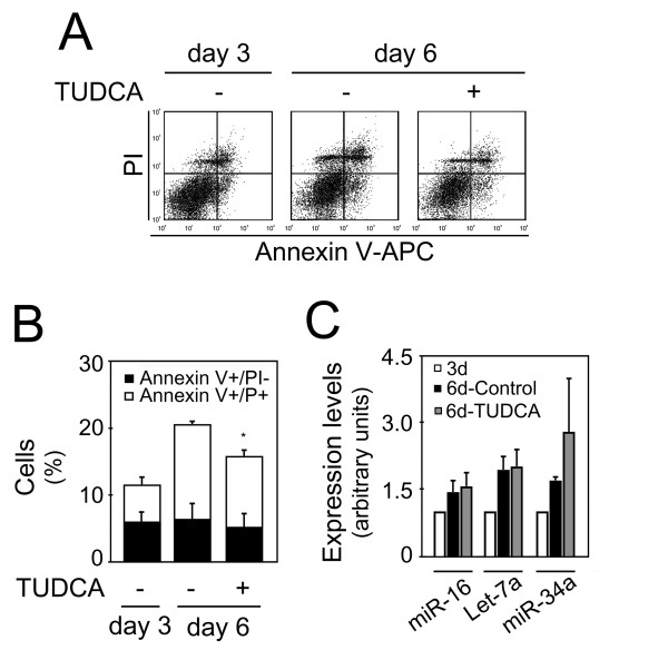
Inhibition of apoptosis by TUDCA was not associated with a decrease in proapoptotic miRNAs expression. Mouse NS cells with 3 days of differentiation were either untreated or treated with 50 μM of TUDCA for 72 hours. Collected cells were stained with Annexin-V-APC/PI to evaluate cell death, or processed for total RNA extraction and miRNAs expression evaluation by quantitative Real Time-PCR. A) Representative Annexin V-APC/PI stainings showing decreased cell death after TUDCA incubation. B) Quantitation of either dying (Annexin+/PI-) or dead (Annexin+/PI+) cells depicted in FACS diagrams. Results are mean ± SEM of triplicates. C) Expression of proapoptotic miRNAs at 3 and 6 days, with or without TUDCA treatment. miR-16, let-7a and miR-34a expression were evaluated from 10 ng of total RNA, using specific primers for each miRNA, and GAPDH for normalization. Expression levels were calculated by the ΔΔCt method using differentiated cells at 3 days as calibrator. Data represent mean ± SEM of three independent experiments. *p < 0.05 compared to respective nontreated cells at 6 days.

