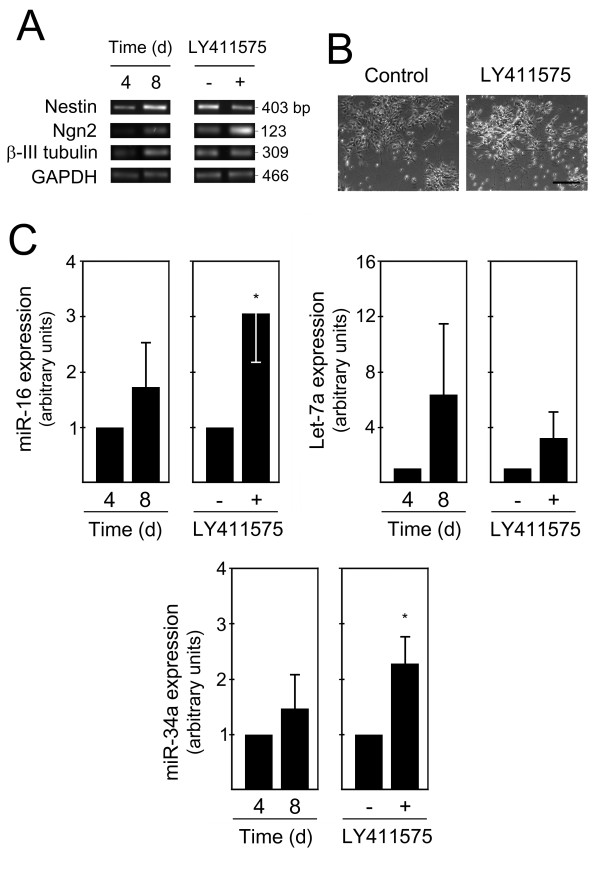
Figure 5.
miR-16, let-7a and miR-34a are increased during mouse ES cell differentiation. Mouse ES cells (Sox1-GFP 46C) were differentiated using an adherent monolayer protocol. Cells at 4 and 8 days were collected for total RNA extraction and subsequently processed for evaluation of specific differentiation markers, as well as proapoptotic miRNA expression by quantitative Real Time-PCR. A positive control for neural differentiation was also performed at day 8 by treating cells with either 10 nM LY411575 or 0.01%DMSO (control) for 12 hours. A) Semi-quantitative RT-PCR analysis for selected markers of lineage commitment in day 4 and 8, as well as in LY411575-treated and untreated cells. B) Representative bright-field, phase contrast images showing increased neuronal differentiation after LY411575 incubation compared with control (DMSO-treated) cells. C) Expression of miR-16, Let-7a and miR-34a at 4 and 8 days of ES cell differentiation and in control (DMSO-treated) and LY411575-treated rosette cultures at 8 days. miRNAs expression were evaluated from 10 ng of total RNA, using specific primers for each miRNA, and GAPDH for normalization. Expression levels were calculated by the ΔΔCt method using either differentiated cells at 4 days or LY411575-untreated cells as calibrator. Data represent mean ± SEM of three independent experiments. *p < 0.05 compared to respective nontreated cells. Scale bar: 50 μm. d, days.

