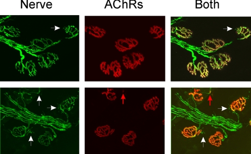
Fig. 6.
Increase in n following block of acetylcholine receptors is not due to new synaptic sites. Shown in each row is a set of endplates from muscle injected with BTX 4 days earlier. Nerve staining is shown in the first column in green, acetylcholine receptors stained with rhodamine-conjugated BTX are shown in red in the second column, and the superimposed images are shown in the third column. In both sets of endplates small sprouts can be seen (white arrows). However, only one of the sprouts is associated with a small region of acetylcholine receptors (red arrow).

