Fig. 3.
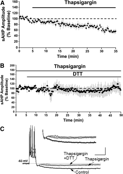
Intracellular Ca2+ stores underlie DTT-mediated decrease in aged-sAHP. (A) Time course of the change in the normalized sAHP amplitude in the aged animals on application of thapsigargin (n = 7). (B) Time course of change in the normalized sAHP amplitude in cells (n = 4) incubated with thapsigargin prior to and during DTT application. (C) Representative traces illustrating the AHP of aged animals under control condition (black trace), and at the end of 40 min application of thapsigargin (gray trace) and at the end of 50 min application of thapsigargin+DTT (gray trace). Calibration bars: 200 ms, 10 mV. Inset: Magnified representation of change in the aged AHP under control condition, thapsigargin, and thapsigargin + DTT.

