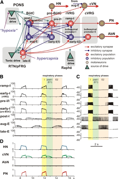
Fig. 7.
The extended model of the brain stem respiratory network. A: schematic of the extended model showing interactions between different populations of respiratory neurons within major brain stem compartments involved in the control of breathing (pons, RTN/pFRG, BötC, pre-BötC, rVRG, and cVRG). Each population (shown as a sphere) consists of 50 single-compartment neurons described in the Hodgkin-Huxley style. In comparison with the previous model (Smith et al. 2007), this model additionally incorporates the population of bulbospinal premotor expiratory (E) neurons in cVRG, representing the source of AbN activity, and the late-E population in the RTN/pFRG compartment (see text for details), serving as a source of RTN/pFRG oscillations. Justification for all interconnections used in the basic models can be found (with the corresponding references) in our previous papers (Rubin et al. 2009; Rybak et al. 2007; Smith et al. 2007). Justification of interconnections involving the late-E population is in the text. The model includes 3 sources of tonic excitatory drive: pons, RTN, and raphé—all shown as green triangles. These drives, especially those from the pontine and RTN sources project to multiple neural populations in the model (green arrows). However, to simplify the schematic, only the most important connections are shown connected to particular populations. The full structure of connections from each drive source [drive(pons); drive(RTN); drive(raphé)] to target neural populations of the model and the corresponding synaptic weights can be found in Table 1. The late-E population receives an additional external drive simulating the effect of hypercapnia; the pontine drive is considered to be hypoxia/anoxia dependent and was reduced in simulation of hypoxic conditions. B: model performance under normal conditions. The activity of major neural populations in the model are represented by average histograms of activity of all neurons in each population (bin = 30 ms). The shown populations include (top-down): ramp-inspiratory (ramp-I located in rVRG), early-inspiratory [early-I(2) in rVRG], preinspiratory/inspiratory (pre-I/I in pre-BötC), early-inspiratory [early-I(1) in pre-BötC], postinspiratory (post-I in BötC), augmenting expiratory (aug-E in BötC), and late-expiratory (late-E in RTN/pFRG). The latter population is silent under normal conditions. C: traces of membrane potentials of the corresponding single neurons (randomly selected from each population). D: the dynamics of the model's motor outputs: HN (blue); cVN (green); AbN (black, silent under normal conditions); PN (red). In B–D, the 3 phase of respiratory cycle are highlighted: inspiratory (I, yellow), postinspiratory (post-I or E1, light green), second expiratory (E2, pink). It is seen that pre-I/I neurons and HN start firing in advance of the beginning of inspiration defined by the onset of PN (and the ramp-I population).

