Fig. 6.
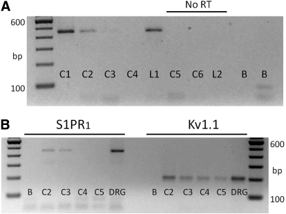
Single-cell RT-PCR analysis shows that some, but not all, small diameter sensory neurons express the mRNA for S1PR1. A: representative gel for the detection of S1PR1 in 5 individual sensory neurons. The lanes labeled C1–C6 (cell1–cell6) are the mRNAs from individual small diameter neurons, whereas those labeled L1–L2 (large cell1 and cell2) are mRNAs obtained from individual large diameter neurons. Lanes C1–L1 represent the detection of S1PR1 amplicons with a product size of 448 bp. Lanes C5–L2 underwent PCR in the absence of RT (No RT). Lanes labeled B (blank) represent reactions performed in the absence of any template. B: in a different tissue harvest than shown in A, the detection of S1PR1 and the potassium channel, Kv1.1 from the same 4 individual sensory neurons. For small diameter sensory neurons C2–C5, Kv1.1 amplicons were detected, whereas S1PR1 was detected in only 2 of the 4 neurons. The lanes labeled DRG were amplicons obtained from mRNA isolated from 10 ganglia.

