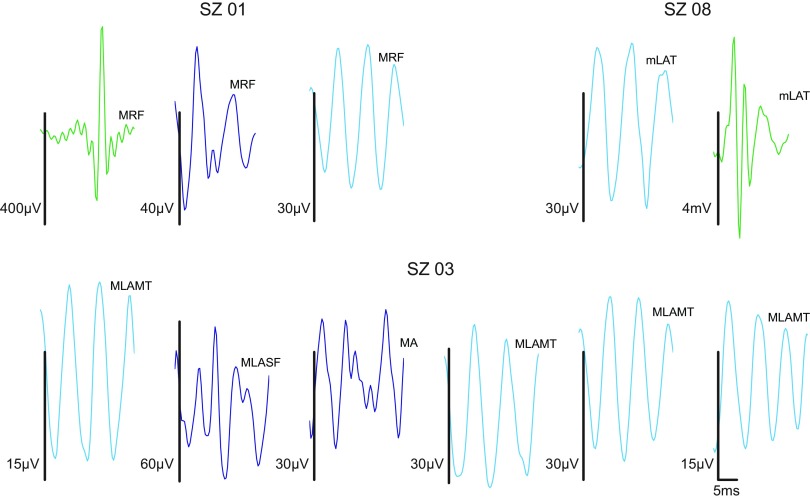
Fig. 7.
Example prototypes from individual patient clustering; 100–500 Hz band-passed waveforms only. Vertical scale bars are 35 μV; horizontal scale bar is 5 ms [to facilitate comparison, events are plotted on identical time axes (0 to 25 ms; axes suppressed in individual plots), with events truncated to 25 ms if longer than this duration]. Subjects SZ01 and SZ08 show clusters that appear to be subsets of the 4 clusters found in the aggregate. Subject SZ03 does as well, but in this case it appears that 2 groups have been oversplit by the algorithm, yielding 6 clusters. The latter type of cluster stability issue was not seen in the aggregate clustering. In the abbreviations above each trace, the first letter indicates whether the recording comes from a macro- (M) or microelectrode (m). RF, right frontal; LAMT, left anterior mesial temporal; A, anterior; LASF, left anterior subfrontal; LAT, left anterior temporal. Note that identical labels do not imply identical electrodes, only that the electrodes have the same lobar location.

