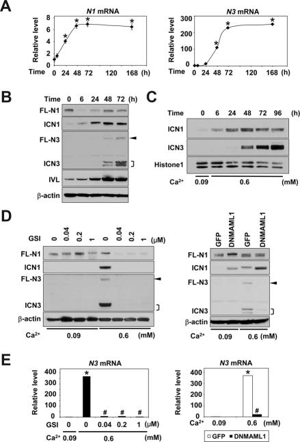
Figure 4.
Sequential induction and activation of N1 and N3 during Ca2+-induced squamous differentiation in esophageal keratinocytes
EPC2-hTERT cells were exposed to 0.6 mM Ca2+ for indicated time periods in (A)–(C) or 72 hrs in (D) and (E), and subjected to real-time RT-PCR in (A) and (E) or Western blotting in (B)–(D). In (D) and (E), Ca2+ stimulation was done in the presence or absence of GSI or DNMAML1. Compound E (GSI) was used at indicated concentrations. β-actin served as an internal or loading control in (A), (B), (D) and (E). Histone H1 served as a loading control for nuclear extracts in (C). *, P< 0.001 vs. time 0 (n=3) in (A). *, P< 0.001 vs. 0.09 mM Ca2+ + 0 μM GSI or GFP; #, P< 0.001 vs. 0.6 mM Ca2+ + 0 μM GSI or GFP (n=3) in (E). FL-N, full-length Notch; ICN, intracellular Notch. Arrowhead, FL-N3;], doublets of ICN3.

