Figure 3.
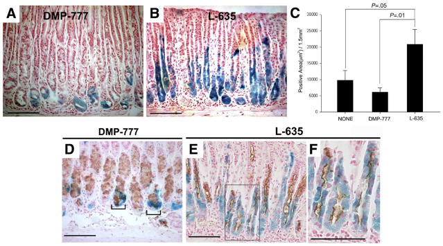
Mist1CreER/+/Rosa26RLacZ mice treated with DMP-777 or L-635. (A and B) Mist1 cell lineage analysis (X-gal staining) after treatment with either (A) DMP-777 or (B) L-635 for inducing SPEM. (C) β-galactosidase–expressing cells were quantitated as an X-gal–stained area per 1.5 mm2 of total mucosal tissue (±standard error of the mean). Compared with both untreated animals (n = 4) and DMP-777–treated mice (n = 8), L-635 treatment (n = 6) caused a significant expansion of the number of X-gal–stained cells. (D–F) Immunostaining for TFF2 in X-gal–stained sections from DMP-777 and L-635–treated mice. Brown (3,3-diaminobenzidine) = TFF2. (D) DMP-777 induced a marked loss of parietal cells and prominent SPEM, with co-staining for X-gal and TFF2 at the bases of glands (brackets). (E) TFF2 expression was observed throughout the X-gal–stained cells in mice treated with L-635. (F) Higher magnification view of panel E. Bar, 100 μm.

