
Figure 4.
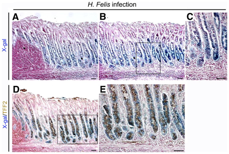
Lineage mapping of SPEM in 6-month-old H felis–infected Mist1CreER/+/Rosa26RLacZ mice. Sections of fundic mucosa stained with (A–C) X-gal or dual stained for (D and E) X-gal and TFF2 (brown). Chronic H felis infection caused an expansion of the number of X-gal–stained cells. (A) Note the lymphoid follicle and inflammatory cell infiltration in the mucosa. (B) Mucosa adjacent to section in panel A. (C) Higher magnification view of panel B. (D) Dual immunostaining for TFF2 in X-gal–stained sections. Brown (3,3-diaminobenzidine) = TFF2. TFF2 expression was observed throughout the X-gal–stained cells in H felis–infected mice. (E) Higher magnification view of panel D. Bar, 50 μm.