
Figure 3. FACS-isolated CD166 epithelial cells express Paneth cell granules or Lgr5.
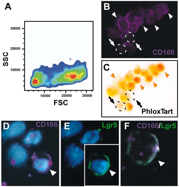
(A) Forward (FSC) and side (SSC) scatter analysis of CD166-expressing crypt epithelial cells display two distinct populations of cells (orange centers). (B-C) FACS-isolated, cytospun CD166-expressing mouse crypt cells (B; purple) stained with (C) Phloxine Tartrazine. White arrowheads designate CD166-positive cells; orange arrowheads designate CD166-positive, Phloxine Tartrazine-positive cells. Arrows and dashed circles designate CD166-positive, Phloxine Tartrazine-negative cells. (D-F) Cytospun, isolated CD166-positive cells (purple) co-stained with antibodies to Lgr5, a putative stem cell marker (green). Arrowhead designates a double-labeled cell.