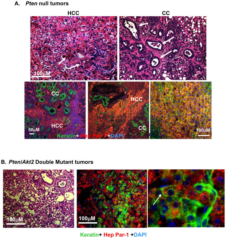
Figure 5. Deletion of Akt2 does not alter the mixed cell tumor phenotype in liver specific Pten null mice.
A. Top, H&E images of tumors developed in the Pten null mice demonstrate compact trabecular growth patterns and pseudoglandular structures of HCC (arrows, left panel); and tubular features of CC (arrows, right panel). Bottom, Immunohistochemistry of liver tissue with HepPar-1 (red) and keratin (green) in Pm mice identifies HCC and CC respectively. Blue, DAPI. B. H&E section of one of the two tumors formed in the Pten/Akt2 double mutant liver (left panel). Immunochemical staining confirms bilineage tumor development with both Hep Par-1 (red) and keratin (green) (right two panels). Arrow point to bilineage cells expressing both markers.

