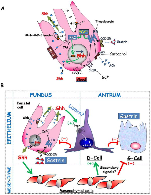
Figure 7. Hypothetical model of Shh in the gastric mucosa.
A) Shown are three mechanisms capable of stimulating acid secretion through an increase in Ca2+i released from the endoplasmic and sarcoplasmic reticulae: 1) hormonal-stimulation with gastrin, 2) cholinergic stimulation with acetylecholine (ACh) through the M3 muscarinic receptor, or 3) alkalinization of the basolateral surface by extruded HCO3− ions leading to the activation of the calcium-sensing receptor (CaSR). Stimulation of acid secretion by histamine release from enterochromaffin-like cells is not shown. Ca2+i-release stimulates acid secretion and Shh gene expression via PKC α and β. PKC-δ might also mediate diacylglycerol (DAG)-induction of Shh gene expression. DAG also stimulates Ca2+i-release and acid secretion. The compounds used in this study, gadolinium (Gd3+), carbachol, thapsigargin and TPA, all stimulated Shh gene expression and are known to increase Ca2+i. Shh protein undergoes processing (intracellular location not defined), migrates to the basolateral membrane, or co-migrates with the H+/K+-ATPase-β subunit to the apical membrane where it is likely to be secreted luminally. B) Luminal Shh potentially targets epithelial cells expressing Ptch-1 such as D-cells to induce non-canonical signaling. In addition, Shh protein reaching the basolateral membrane would target Gli-1-positive mesenchymal cells inducing a secondary signal from the mesenchyme. The outcome of Shh signaling is to induce somatostatin (SST) that in turn inhibits the G-cell and gastrin production, and acid secretion from the parietal cell through paracrine mechanisms.

