Figure 1a.

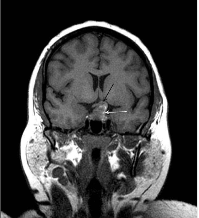
Coronal T1W MR image shows a predominantly isointense mass with few hyperintense areas (white arrow). It is bulging to left of midline and causing compression of left half of optic chiasma (black arrow)

Coronal T1W MR image shows a predominantly isointense mass with few hyperintense areas (white arrow). It is bulging to left of midline and causing compression of left half of optic chiasma (black arrow)