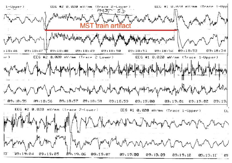
Figure 5. Scalp EEG of a patient undergoing high dose MST for treatment of depression.

This is a representative 2-channel, bifrontotemporal scalp EEG recording during one of the initial human HD-MST sessions from the case series reported in Kirov et al. (Kirov, et al., 2008). The period of the MST train is marked. Following the train, high frequency spiking can be seen in both channels that evolves into spike-slow wave configuration, and then slow wave activity. This was accompanied by a generalized tonic-clonic seizure as documented by motor convulsion in an unanesthetized limb, employing the cuff-technique.
