Fig. 6.
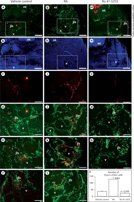
In utero exposure of mouse embryos to excess of RA increases the number of Prox1-positive lymphatic progenitor cells in the cardinal veins and jugular lymph sac forming areas. Double-immunofluorescence analysis of ED 11.5 mouse embryos exposed to excess RA (g–l) or vehicle (a–f) for CD31 (cell membrane staining) and Prox1 (cell nucleus staining) revealed that RA exposure increased the number of Prox1+ endothelial cells in the anterior cardinal veins and in the area of the jugular lymph sacs as compared with control (r). Ro 41-5253 treatment (m–q) slightly reduced the number of Prox1+/CD31+ cells in the jugular area as compared with control (r). d–f, j–l and p, q show representative sections of embryos from independent vehicle control, RA- or Ro 41-5253-treated pregnant mice, respectively. Cell nuclei are shown in b, h, n. a, b, g, h, m, n Scale bars = 100 μm; c–f, i–l, o–q scale bars = 50 μm. a = Dorsal aorta; jls = jugular lymph sac; nt = neural tube; v = cardinal vein. Data are expressed as mean values + SD (n = 4). *** p < 0.001.

