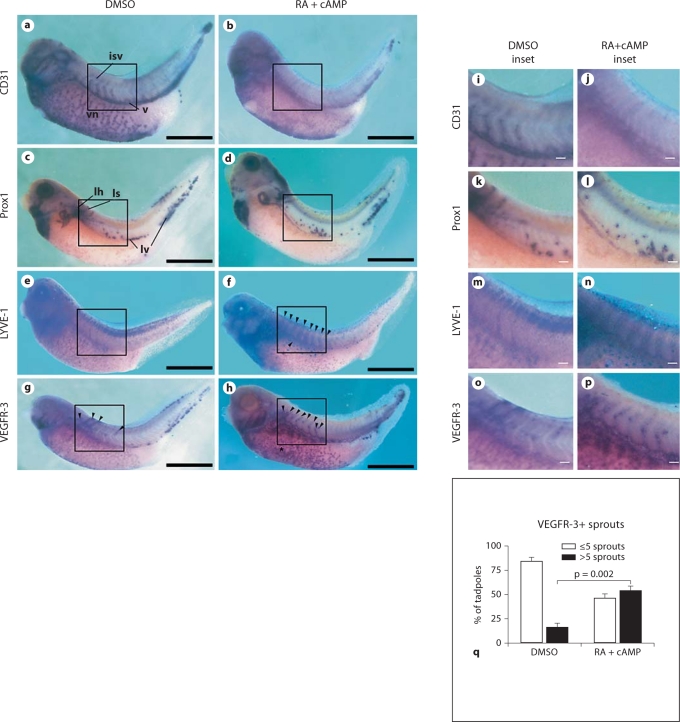
Fig. 7.
Incubation of X. laevis embryos with excess of RA downregulates CD31 and upregulates the lymphatic markers LYVE-1 and VEGFR-3 in the developing vasculature. X. laevis tadpoles were bathed in RA in combination with cAMP (b, d, f, h, j, l, n, p) or in DMSO (a, c, e, g, i, k, m, o) from embryonic stages 28 to 39. In situ hybridization for CD31 (a, b, i, j), Prox1 (c, d, k, l), LYVE-1 (e, f, m, n) or VEGFR-3 (g, h, o, p) revealed that RA and cAMP reduced CD31 expression (b, j) in the developing vasculature, including intersomitic veins (isv), the cardinal vein (v) and the vitelline network (vn), compared with controls (a, i). No effects on Prox1 expression and/or distribution in the lymph heart (lh), lymphatic sprouts (ls) or in the lymphatic endothelial cells of venous origin (lv) were observed (c, d, k, l). In contrast, LYVE-1 expression was upregulated in the intersomitic veins and in the cardinal vein (f, arrowheads, n) compared with controls (e, m). RA incubation also upregulated VEGFR-3 expression in the cardinal vein and vitelline network (h, arrowheads and asterisk, respectively, p) compared with control embryos (g, arrowheads, o). Scale bars = 50 μm; i–p scale bars = 5 μm. q Quantitative analysis of the number of VEGFR-3+ sprouts from the anterior cardinal vein following exposure to DMSO (vehicle) or the combination of RA and cAMP. Data are expressed as means + SEM (n = 150 per treatment group).

