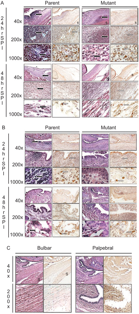
FIGURE 4.
Representative histology pictures of (A) bulbar conjunctivae or (B) palpebral conjunctivae that were infected with parent strain K1544 and mutant strain K1544ΔCAP at 24 and 48 hr post-infection. A non-infected negative control (C) is also shown. The first column at each time point is stained with hematoxylin and eosin while the second column is stained with a mouse anti-rabbit monoclonal antibody specific for macrophages and granulocytes. Black arrow indicates PMN. White arrow indicates macrophage. S: sclera; R: retina.

