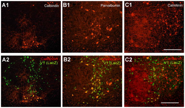
Figure 4. Expression of calbindin, parvalbumin or calretinin in V1-derived interneurons.
A, Ventral horn dual-immunolabeled for calbindin and βgal shown with calbindin-immunoreactivity only (A1) and βgal superimposed (A2). B, C, Similar images for parvalbumin and βgal (B1,2) and calretinin and βgal (C1,2). All ventral calbindin-IR neurons (group 1; Renshaw cells) were βgal-IR. Similarly, most parvalbumin-IR neurons close to LIX (groups 1 and 4) were also βgal-IR. Dorsomedial calbindin-IR neurons (group 3), most calretinin neurons (groups 6 and 7) and ventromedial parvalbumin neurons (groups 5 and 6) were almost never βgal-IR. Around half of calbindin-IR neurons located in mid-regions of the ventral horn (group 2) were βgal-IR. Quantitative data from these preparations are shown in Figure 5. Scale Bars in C1 and C2, 200 μm.

