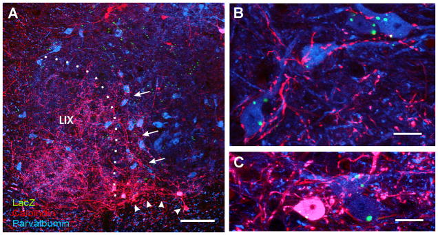
Figure 6. Ia inhibitory interneurons are derived from V1-interneurons.
A, Low magnification image showing parvalbumin-IR neurons in blue (Cy5), calbindin-IR neurons in red (Cy3) and βgal-immunoreactivity in green (FITC). Almost all ventrally located neurons in red (calbindin only) or pink (calbindin + parvalbumin co-expression) are V1-derived (arrowheads) and correspond to Renshaw cells (group 1). Dorsally located calbindin-IR neurons (red) do not co-express parvalbumin and are not V1-derived. Blue neurons (parvalbumin-only) in close spatial relationship to LIX (group 4) are V1 derived, while more medially located parvalbumin-IR neuron are not. A dense plexus of calbindin-IR fibers extends from the Renshaw cell area into LIX and also into the LVII region that contains many V1-derived parvalbumin-IR neurons (arrows). B, High magnification of mid LVII showing V1-derived (βgal-IR) parvalbumin-IR neurons (blue) receiving a dense innervation from calbindin-IR fibers (red). C, A similar image to B, but from ventral LVII and including one Renshaw cell co-expressing both calbindin and parvalbumin (pink). Calbindin-IR contacts were more numerous on V1-derived parvalbumin-only neurons than on Renshaw cells or other neurons that lacked parvalbumin or calbindin (immunostained in other preparations with NeuN, not shown). Almost all neurons densely innervated by calbindin-IR fibers were parvalbumin-IR and V1-derived. These neurons likely correspond with Ia inhibitory interneurons (see text and Figure 2). B and C are superimpositions of 3 confocal planes separated by 1 μm. Scale bars: A, 100 μm; B,C, 10 μm.

