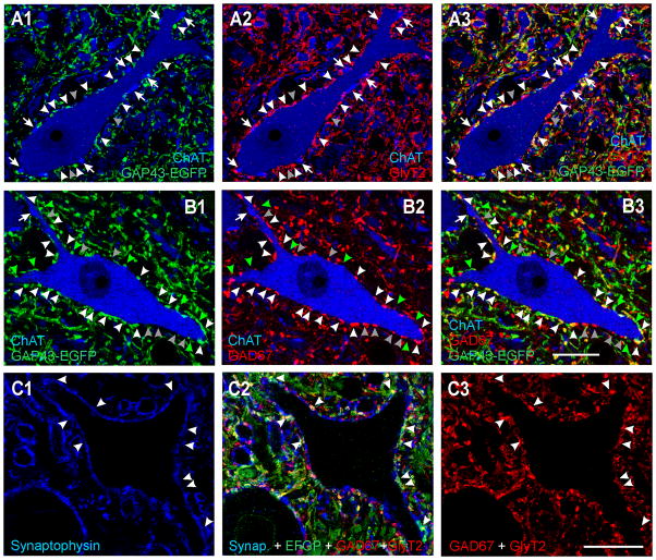
Figure 8. V1 axons express markers indicating a glycinergic and/or GABA phenotype.
A, High magnification single confocal optical section through a motoneuron cell body (ChAT-IR, blue, Cy5) surrounded by V1-derived axons (GAP43-EGFP, green, Alexa 488 in A1) and GlyT2-IR axons (Cy3, red, A2). Both images are superimposed in E3. White arrowheads indicate V1-varicosities that also contain GlyT2-immunoreactivity and are in contact with the motoneuron. Grey arrowheads indicate some GlyT2-IR varicosities in contact with the motoneuron that are not V1-derived (GAP43-EGFP negative). Arrows point to ChAT-IR C-terminals, none of which were V1-derived. Most V1-derived contacts on motoneurons express GlyT2-IR suggesting a glycinergic phenotype. B, High magnification single confocal optical section through another motoneuron in a section stained for GAD67-immunoreactivity. A proportion of V1 derived varicosities were GAD67-IR (white arrowheads) but there were also significant numbers of GAD67-IR terminals that were not V1-derived (grey arrowheads) and many V1-derived varicosities that did not contain GAD67-IR (green arrowheads). ChAT-IR C-terminals (one marked with an arrow) were not V1-derived or GAD67-IR. C, A region of lamina IX neuropil with unstained motoeneuron cell bodies and proximal dendrites and triple immunolabeled for synaptophysin (blue, Cy5), EGFP V1 axons (Alexa 488, green) and GAD67-GlyT2, both antibodies detected with the same fluorochrome (TRITC, red). C1 shows synaptophysin immunolabeling alone. C2, EGFP and GAD67/GlyT2 immunostaining superimposed. C3, GAD67/GlyT2 immunoreactivities. Note that some boutons are filled with immunoreactivity due to intense GAD67 and others are only labeled in the periphery, as characteristic for GlyT2. Arrowheads are in the same positions in C1, C2, and C3 and indicate V1 EGFP positive varicosities containing synaptophysin (i.e., synaptic sites). All V1 synaptic sites contain some red immunofluorescence indicating that they are GAD67 and/or GlyT2 immunoreactive and therefore inhibitory. Similar analyses were performed in random regions of LIX and LVII neuropil with similar results (not shown in figure). Scale bars, B3, C3, 20 μm. Panels A nad B are at the same magnification.

