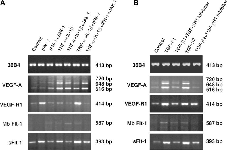
Fig. 4.
RT-PCR analysis of VEGF-A, VEGF-R1 (Flt-1), MbFlt-1, and sFlt-1 mRNA in HCRF cells. (A) Effects of IFN-γ (100 U/ml), TNF-α (10 ng/ml), IL-1β (10 ng/ml), and JAK-1 inhibitor (1 μM). HCRF cultures were incubated in serum free medium for 8 h in the presence of various combinations as indicated in the figure. Total RNA prepared was used for RT-PCR as described in the Methods section. (B) Effect of TGF-β1(10 ng/ml), TGF-β2 (10 ng/ml) and TGF-βR1 inhibitor (100 nM). HCRF cultures were incubated in serum free medium for 8 h in the presence of various combinations as indicated in the figure and RT-PCR performed as described above. PCR reactions in (A) and (B) were performed for 25 (36B4) or 30 (all others) cycles. Each panel is amplified to different levels to visualize the bands; therefore, comparisons may not be made between the panels.

