Figure 3.
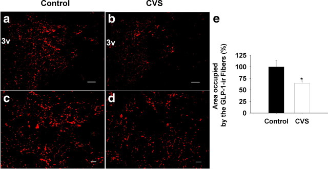
Quantification of the expression of GLP-1 fiber in the mpPVN after 2 weeks of CVS exposure. The field area occupied by GLP-1-immunoreactive positive fibers was determined and expressed as a percentage of total measured area (FldAreaP). Data are shown as percentage of control. a, b, Representative images for low-magnification GLP-1 fiber staining in the PVN. mpPVN, Medial parvocellular PVN; 3v, third ventricle. c, d, Representative images for high magnification of projection images. Left, Control; right, CVS. e, Quantification of the expression of GLP-1 fiber in the mpPVN after CVS exposure. The percentage of area occupied by GLP-1-positive fibers declined after 2 weeks of CVS exposure compared with the control. Scale bars: a, b, 100 μm; c, d, 20 μm. *p < 0.05 versus control.

