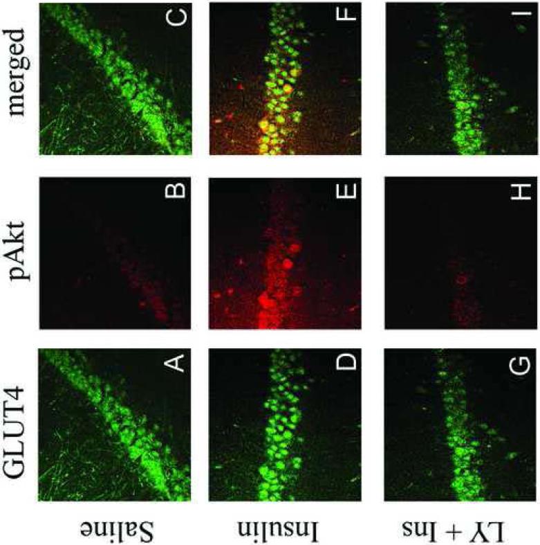
Figure 10.
Insulin stimulated phosphorylation of Akt is colocalized with GLUT4 in the rat hippocampus. GLUT4 (green immunofluorescence) exhibits the expected immunofluorescence distribution in the CA3 region of the rat hippocampus in saline-treated rats (Panel A), insulin-treated rats (Panel D) and rats pre-treated with the PI3-kinase inhibitor LY294002 prior to insulin treatment (Panel G). Insulin treatment increases pAkt immunofluorescence in the CA3 region of the hippocampus (Panel E; red fluorescence), increases not observed in rats pre-treated with LY294002 (Panel H). The phosphorylated form of Akt is not detected in saline-treated rats (Panel B). The merged images illustrate that pAkt exhibits colocalization with GLUT4 in the hippocampus of insulin-treated rats (Panel F).

