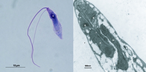
FIG. 1.—
Structure of Proteromonas lacertae. Left panel: Light micrograph of P. lacertae. The Giemsa-stained cell has a size of ∼13 × 3 μm. The anterior of the cell bears two flagella, one thicker and longer than the other. The single nucleus (n) is visible at the anterior pole of the cell. Right panel: Transmission electron micrograph of P. lacertae. This section of the anterior of the cell shows the rhizoplast (r) passing through the Golgi apparatus (g) and into the nucleus (n). The single large mitochondrion (m), in which cristae are visible, is adjacent to the nucleus. For a more detailed description see Brugerolle and Mignot (1990).

