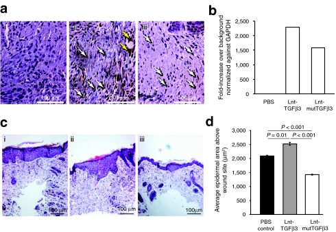
Figure 5.
Mice were treated with a single intradermal injection of 1 × 10⁹ vp of lentivirus expressing either TGFβ3, mutTGFβ3, or a sham PBS injection. This was followed by a full-thickness midline incision of the skin over the injection site 4 hours later. After 14 days, the animals were sacrificed and the incision site was dissected out and analyzed by immunohistochemistry and real-time qPCR. (a) Sections were stained for GFP (i) PBS, (ii) Lnt-TGFβ3, (iii) Lnt-mutTGFβ3 which showed GFP positive fibroblasts (white arrow) and other cell types such as endothelium (yellow arrows). Images are representative for each treatment (n = 6). (b) Relative levels of GFP were quantified using qPCR with an n of three samples carried out duplicate. (c) H&E stained sections were analyzed for re-epithelialization area at the site of incision (i) PBS, (ii) Lnt-TGFβ3, and Lnt-mutTGFβ3. (d) Quantification of re-epithelialization using Image J analysis software of at least 10 sections cut at 50 micron intervals across the wound site for each treatment group and expressed in microns sq. (n = 4, 3, 4, respectively). mutTGFβ3, mutant transforming growth factor β3; PBS, phosphate-buffered saline; qPCR, quantitative PCR; vp, virus particles.

