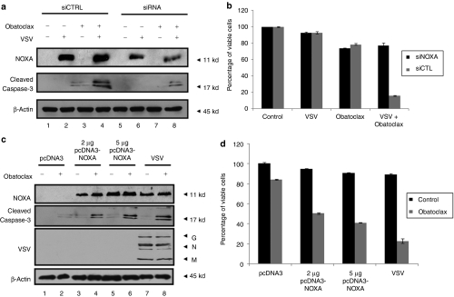
Figure 4.
Induction of NOXA expression is necessary and sufficient to synergistically induce apoptosis with obatoclax. (a–b) Karpas-422 B-lymphoma cells were transiently transfected with siRNA targeting human NOXA (siNOXA) or the nontargeting control pool (siControl). At 24 hours post-transfection, cells were treated with obatoclax followed or not by VSV infection. (a) At 24 hours postinfection, cells were lysed and NOXA silencing was analyzed by immunoblot using anti-NOXA antibody. Caspase-3 cleavage was also determined using an anti-caspase-3 antibody. (b) Cell viability analysis by Annexin V/PI staining was performed on cells treated as described in a. Black bars represent Karpas-422 cells treated with siNOXA and gray bars represent cells treated with siControl. The data shown are the mean ± SEM (n = 3). (c–d) Karpas-422 B-lymphoma cells were transiently transfected with human pcDNA3-NOXA or empty vector. At 24 hours post-transfection, cells were treated with obatoclax (a). At 24 hours post-treatment, cells were lysed and NOXA expression, caspase-3 cleavage and VSV replication were analyzed by immunoblot. G, glycoprotein; M, matrix; N, nucleocapsid. (b) Cell viability was determined by FACS analysis after Annexin V/PI staining. (d) Black bars represent nontreated Karpas-422 cells and gray bars represent cells treated with obatoclax. The data shown are the mean ± SEM (n = 3). FACS, fluorescence-activated cell sorting; VSV, vesicular stomatitis virus.

