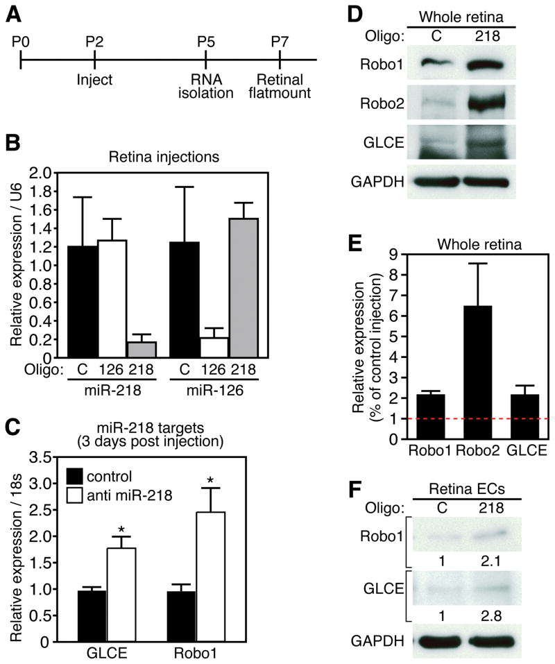
Figure 5. miR-218 modulates expression of Slit-Robo axis genes in the retina.
A. Experimental setup of retinal injections. Postnatal (P) age is denoted above line. Timepoints of injection and sample isolation are denoted below line. B. Real Time RT-PCR demonstrates knockdown of miR-218 or miR-126 in retinal samples 3 days after injection with LNA modified antisense miR-218 or miR-126 oligonucleotides. Expression is relative to control oligonucleotide and normalized to U6. C. Real time RT-PCR reveals expression levels of GLCE and Robo1 from retinal samples 3-days after injection. Error bars are SEM (n = 3, * denotes p < 0.05). D. Western for Robo1, Robo2, and GLCE from a pool of 5 retinas, 3 days after injection with control or anti miR-218 oligonucleotides. GAPDH is detected as loading control. C = control, 218 = anti miR-218. E. Quantification of protein levels from retinal explant pools. F. Western blot detecting Robo1 and GLCE from retinal ECs 48 hrs after transfection with anti miR-218 oligonucleotides. GAPDH is detected as loading control. Numbers refer to Western band intensity, normalized to GAPDH and relative to control.

