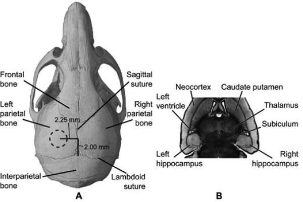
Fig. 2.
Location of FUS-targeted regions in vivo. The FUS transducer was targeted through (A) the left parietal bone of the mouse skull. The dotted circle in (A) approximately corresponds to the 1.32 mm full-width-at-half-maximum peak diameter of the focus. The focal volume was placed to overlap the left hippocampal region as seen on a horizontal histological section (B) of the mouse brain. The right hippocampal region was not targeted and acted as a control.

