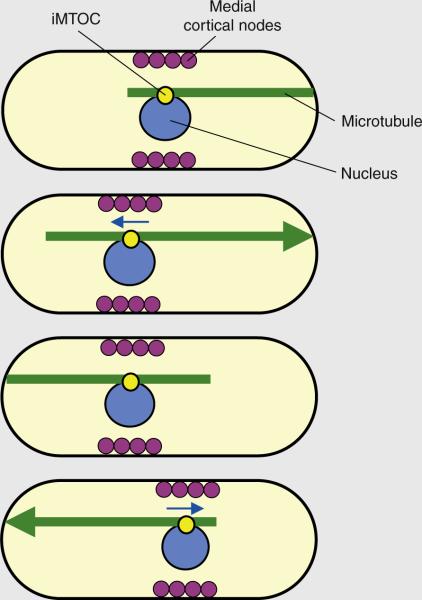
Figure 2.
Microtubule pushing centers the nucleus and cortical medial nodes to define the site of cell division.
The iMTOC tethers the microtubule bundle to the nucleus. During microtubule–cortex contact, sustained polymerization at the microtubule plus end produces a pushing force (shown by the large arrowhead) that displaces the nucleus in the opposite direction (nuclear movement depicted by blue arrows). The antiparallel configuration of the microtubule bundle ensures that, over time, the nucleus oscillates back and forth toward the geometrical center of a growing cell. Coupling between the nucleus and the medial cortical nodes ensures that the nodes, over time, are also positioned at the cell middle. The medial nodes subsequently organize the actomyosin ring for cell division.

