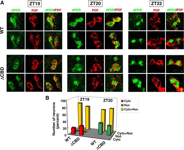
Figure 3.
The timing of dPER nuclear entry in the small ventral lateral neurons of p{dper(ΔCBD)} flies is similar to that observed for p{dper(WT)} flies. Adult flies of the indicated genotypes (left of panel) were collected at the indicated times in an LD cycle and processed for immunohistochemistry followed by visualization using confocal microscopy. A, Shown are representative staining patterns obtained for the small ventral lateral neurons (s-LNvs) from at least 5 flies; dPER was visualized with anti-HA (3F10) antibodies labeled with Alexa Fluor 488 (green). PDF was visualized with anti-PDF (C7) antibodies labeled with Alexa Fluor 533 (Cyran et al., 2005). B, The cytoplasmic/nuclear distribution of dPER for s-LNv from each genotype at ZT19 and ZT20 was quantified.

