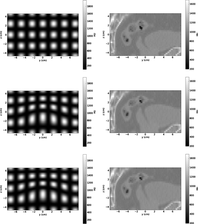
Figure 5.
The region of interest for the source CT images for the numerical phantom (upper left) and the pelvic CT image (upper right), the known deformed CT images for the numerical phantom (middle left) and the pelvic CT image (middle right), and the resultant CT images reconstructed via projection matching for the numerical phantom (lower left) and the pelvic CT image (lower right) show that the source (FBCT) image quality is preserved.

