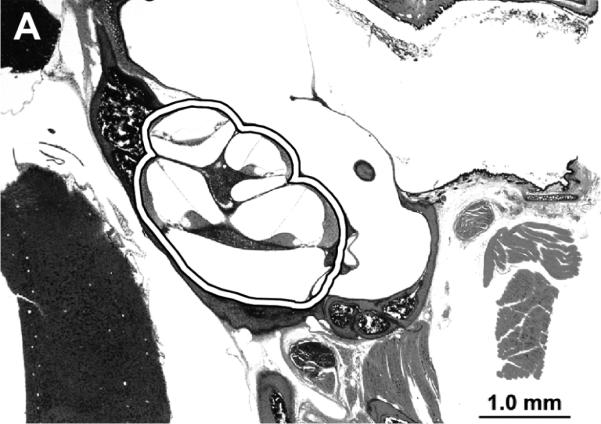
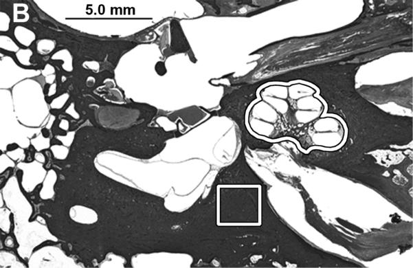
Figure 1.


A) Photomicrograph of mouse celloidin embedded temporal bone section. The sections contained both cochleae and they were removed as outlined for further analysis. B) The cochleae and a sample of the otic capsule bone from cadaveric human celloidin embedded sections were removed (outlined areas). The scale bar is 1.0 mm in A and 5.0 mm in B.
