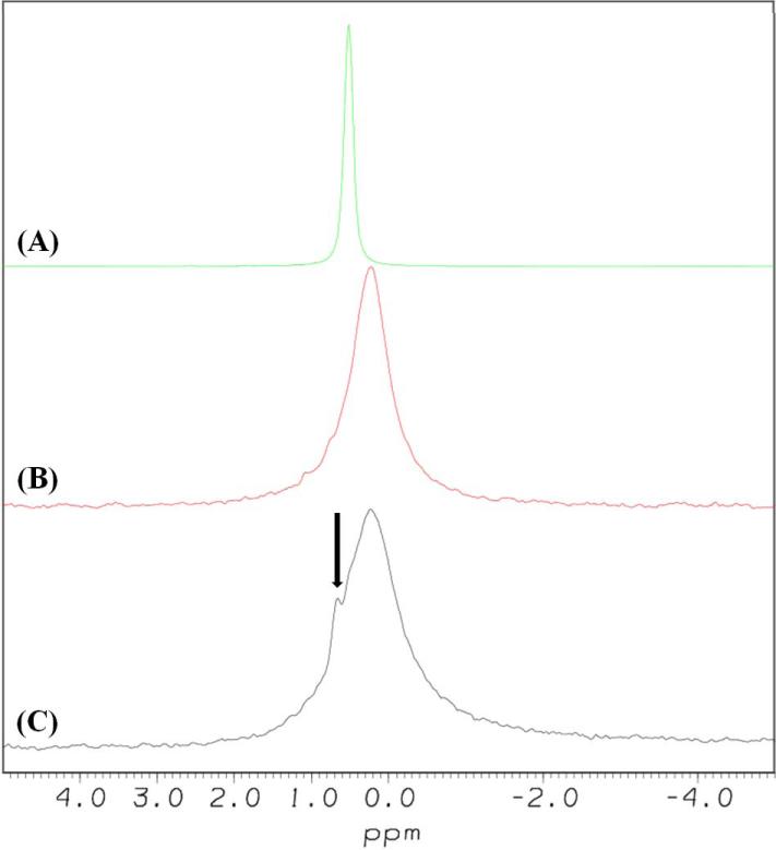
Figure 6. Addition of [113-122]apoJ to KOdiA-PC results in the appearance of an isotropic narrow 31P signal.
31P NMR spectra of DPC micelle (A), KOdiA-PC (B), and mixture of [113-122]apoJ and KOdiA-PC (lipid to peptide molar ratio, 25:1) (C). 2048 scans were used to acquire each 31P spectrum, at a temperature of 310 K. The samples were allowed to equilibrate in the probe for 30 minutes before acquisition was begun. The spectra were processed with exponential multiplier window functions, using a line broadening value of 10 Hz. The spectra were referenced using 85% phosphoric acid (0.0 ppm). The narrow 31P signal, indicated by an arrow (C), presumably arises from the KOdiA-PC complexed with [113-122]apoJ.

