
Figure 3.
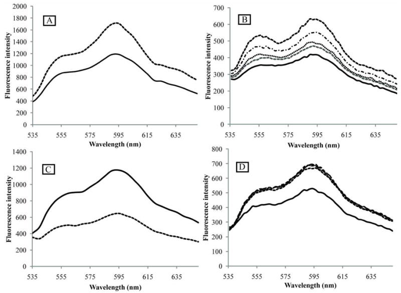
Fluorescence spectra of DOX upon interaction with PEG-PCL block copolymer (A), phospholipid micelles (B), DNA (C), and histone (D). (A) – effect of DOX encapsulation in PEG-PCL micelles. DOX concentration 25 μg/ml (43 μM). Solid line - DOX dissolved in PBS; dashed line – DOX encapsulated in 0.4% PEG-PCL micelles. (B) - effect of PEG-PE micelles. DOX concentration 8.3 μg/ml (14 μM). Solid line – DOX dissolved in PBS; dashed lines – DOX in PEG-PE micelles. PEG-PE concentrations (from bottom to top): 27.8 μg/ml (9.9 μM); 55.6 μg/ml (19.8 μM); 111.2 μg/ml (39.6 μM), and 222.4 μg/ml (79.2 μM). (C) – effect of DOX intercalation into the DNA; DOX concentration 8.3 μg/ml (14 μM). DNA concentration 18.9 μg/ml (30 μM base pairs); Solid line – DOX dissolved in PBS; dashed line – DOX intercalated into the DNA. (D) – effect of DOX interaction with histone. DOX concentration 8.3 μg/ml (14 μM). Solid line – DOX dissolved in PBS; dashed lines - DOX associated with histone; histone concentrations: 1.6 μM; 3.2 μM; 6.4 μM, and 12.8 μM.