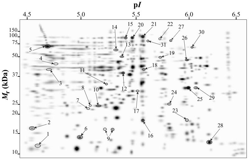
Fig. 2.
PDQuest-generated master gel image showing the general spot pattern of matched spots from TC fractions obtained from A. baumannii ATCC 19606T cells grown under −Fe/−DIP, +Fe/−DIP, −Fe/+DIP or +Fe/+DIP conditions. Differentially expressed spots are numbered. Mr, relative molecular weight; pI, isoelectric point.

