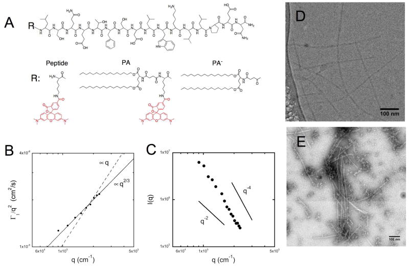
Figure 1. Physicochemical characterization of micelles composed of double-tailed peptide amphiphiles.
Peptide p5314–29 was fluorescently labeled with rhodamine (Peptide) and a synthetic di-palmitic tail (PA). A non-fluorescent peptide amphiphile (PA−) was synthesized to vary micelle fluorescent intensity (A). When dissolved in an aqueous phosphate buffer, PA self-assembled into elongated micelles. Cryogenic (D) and negative stain (E) transmission electron microscopy were used to image the high aspect ratio micelles and dynamic (B) and static (C) light scattering were used to extract information on their size and persistence length.

