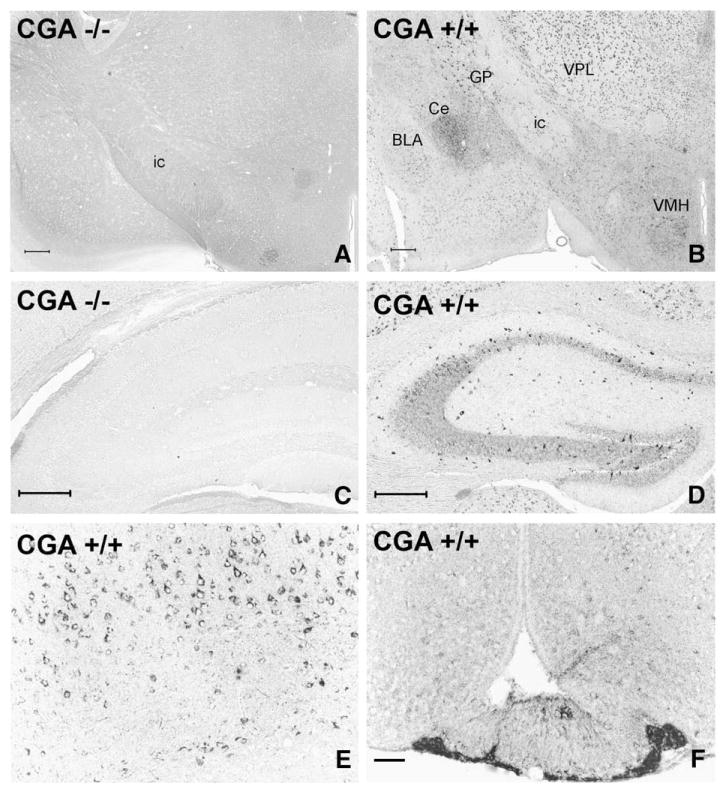
Fig. 1.
Analysis of the specificity and distribution of CGA/WE14 immunostaining in mouse forebrain employing CGA-deficient and wild-type mice. Note total absence of WE14 staining in a coronal brain section at the level of the diencephalon (A) and the hippocampus (C) of a CGA-deficient mouse demonstrating the specificity of WE14 immunostaining for CGA. WE14-immunostaining is present in perikarya of the thalamus, globus pallidus and ventromedial hypothalamus and in fibers of the limbic system of a CGA +/+ mouse (B). Note strong WE14 staining of perikarya of presumed GABAergic interneurons and of fibers in the radial layer of the CA2 and CA3 hippocampal region and in dentate gyrus of a CGA +/+ mouse, while pyramidal and granular cell layers are only weakly WE14-immunopositive (D). The perikarya of layer V pyramidal neurons of parietal neocortex of a CGA +/+ mouse are strongly immunoreactive for CGA (E). Note stronger WE-14 fiber immunostaining in the internal layer of the median eminence (ME) than in the external layer, and weakly WE14-stained neuronal perikarya and fibers in the arcuate nucleus. WE14 immunostaining is strong in the endocrine cells in the tuberal part of the anterior pituitary (F). ic, internal capsule; GP, globus pallidus; VPL, ventral posterolateral thalamic ncl.; Ce, central ncl. of amygdala, BLA, basolateral ncl. of amygdala; VMH, ventromedial hypothalamic ncl. Bars=200 μm (A, B), 100 μm (C, D), 50 μm (E, F).

