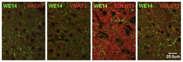
Fig. 10.
Comparative distribution of WE14 immunoreactivity and classical neurotransmitter markers in the rat amygdala. High resolution confocal microscopy analysis of the central ncl. of amygdala after double immunofluorescent staining with WE14 and specific neurotransmitter synaptic markers Note mutual exclusion of WE14 and cholinergic (VAChT) as well as monoaminergic (VMAT2) immunostaining. There is a minor costaining of WE14 with glutamatergic synaptic markers VGluT1 or VGluT2. Bar=20 μm.

