
Fig. 3.
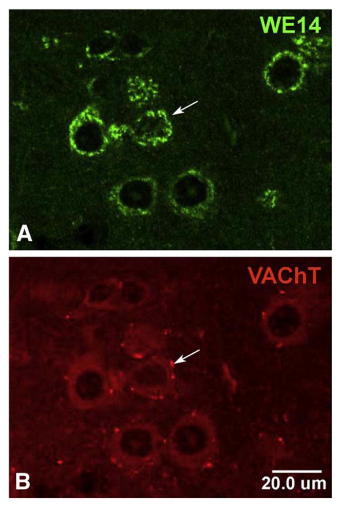
CGA expression in cholinergic motoneurons in mouse spinal cord. Confocal microscopic analysis of motoneurons after double immunofluorescent staining of mouse spinal cord sections with WE14 and VAChT. Staining for WE14 (A) is concentrated in the perikarya of VAChT-positive cholinergic motoneurons (B) but appears to be absent from VAChT-positive cholinergic autapses (arrow). Bar=20 μm.