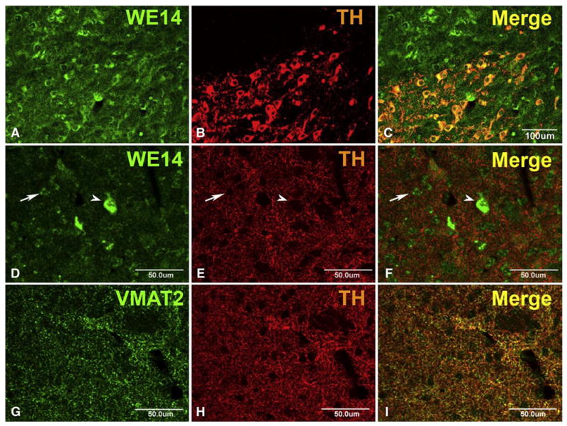
Fig. 6.

CGA expression in mouse nigrostriatal system. Coronal brain sections through the midbrain (A–C) and the striatum (D–F) of an adult CGA +/+ mouse (C57Bl/6). Note preferential perikaryal localization, and similar abundance of WE14 immunoreactivity, in TH-positive neurons of the substantia nigra pars compacta and the adjacent TH-negative nucleus ruber (A–C). WE14 immunostaining is present at high abundance in presumed large cholinergic striatal interneurons (arrow head) and at low abundance in presumed small GABAergic striatal projection neurons (arrow) but virtually absent from TH-positive fibers and terminals in the striatum (D–F). Unlike WE14 staining, VMAT2 staining is present in, and mostly overlapping with, TH-positive varicose fibers in the striatum (G–I). Bar=100 μm (A–C), bar=50 μm (D–I).
