Fig. 1.
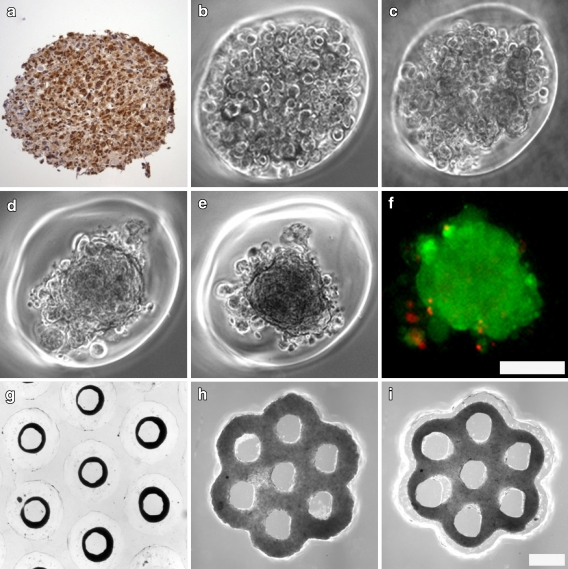
Theca cells can self-assemble into complex 3-dimensional microtissues. Isolation of theca cells from antral follicles was confirmed immunohistochemically with calretinin staining (a). Theca cells were seeded into spheroid shaped wells and self assembly into microtissues was documented starting at 1 h post seeding (b). Theca cells began to aggregate by 4 h (c) and compacted into spheroids by 24 h (d). Compaction into spheroids was complete at 48 h (e) and live-dead staining confirmed the viability (green) of theca cells in complex microtissues (f). Theca cells self-assembled into luminal structures such as toroids (g) seen at 24 h after seeding. Theca cells also formed complex microtissues containing multiple openings (lumina) including the honeycomb at 18 h (i) after initial seeding (h). Scale bars (100 microns) are the same for panels b, c, d, e, f, and for panels g, h,i

