Fig. 4.
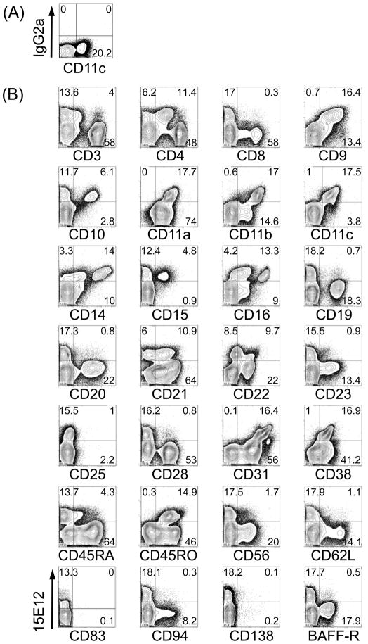
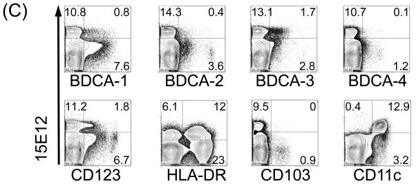
Expression of hDCIR is restricted to monocytes, neutrophiles, granulocytes and dendritic cells. FACS-Density blots show PBMCs that were stained with Alexa-647 labeled (A) isotype control antibody (IgG2a), (B, C) hDCIR antibody clone 15E12 (y-axis) and antibodies characterizing (B) different lymphocytes or (C) DC-subpopulations (x-axis). Shown are living cells. The experiment was repeated three times with different donors.


