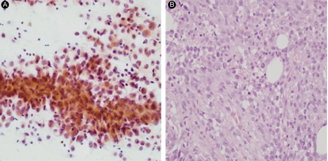
Figure 3.
(A) Fine needle aspiration of the thyroid mass. The tumor is present as loose clusters or single cells and is composed of epithelioid cells with pleomorphic nuclei (Papanicolaou, × 400). (B) Excisional biopsy specimen from the periumbilical area. The tumor is composed of pleomorphic, epithelioid, and spindle cells with frequent mitotic figures (H&E, × 400).

