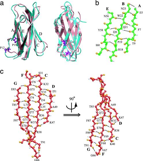
Fig. 2.
Structures of equilibrated FN-III1. (a) Alignment of NMR (cyan) and equilibrated (pink) structures shown in two different orientations. Two prolines that prevent A and B strands forming more interstrand hydrogen bonds are colored purple. Interstrand hydrogen bond networks of the ABE (b) and of the GFCD β-sheets (c), the latter in different orientations. Hydrogen and oxygen atoms are colored white and orange. Hydrogen bonds are represented by black dashed lines.

