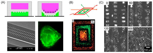
Figure 5.
Substrate patterning using flow or active elements. (A) Structured valve based microfluidic device for substrate patterning. A1—A2: schematics showing cross-sectional view of the device and deformation of structured membrane when applying a pressure on the top channel. Flow of passive agent recovers only the area unprotected by membrane structure, allowing future adhesion of ECM protein. A3: picture shows a scanning electron micrograph of the membrane structure. A4: fluorescence picture of actin stained endothelial cell following ECM matrix protein shape [71]. (B) Direct cell patterning using reversible device with two interconnected channel layers. Up: schematic representation of the method used to pattern different cell types on the same substrate. Down: fluorescence picture of two cell types deposited on a tissue culture dish in a concentric pattern using this device [72]. (C) Dynamic cell patterning. Picture showing BCE cells attached to a surface patterned with specific thiols. Application of a cathode voltage pulse allowed release of the cells from the micropattern (time in minutes) [79].

